|
Diagnostics |
||
|
The presice diagnosis of
filarisis is presently based in the demonstration of the parasites in the
night blood is incovinient, inconsistent and not sensitive in detecting
low microfilaraemia. Further, microfilariae are not
normally seen in peripheral blood in acute and chronic infections
as well as occult filarial infections, making
it difficult to
diagnose clinically for successful treatment.
Most of the clinicians rely on their clinical acumen
in the diagnosis of clinical filariasis.
Immunodiagnosis:
|
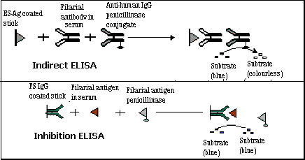
Antibody detection by Indirect Penicillinase ELISA & antigen detection by Inhibition Penicillinase ELISA :The
antibody detection test is based on the detection of specific IgG antibody
to Brugia malayi mf ES antigen by
indirect penicillinase ELISA. The mf ES antigen coated CAM sticks are
incubated with diluted (1:300) filarial sera followed by anti-human IgG
penicillinase conjugate. After washing, when the sticks are finally
incubated with blue coloured starch-iodine-penicillin ‘V‘ substrate, the
disappearance of
the colour earlier than control indicates the presence of filarial
antibody. The free and IC antigen detection is done by inhibition ELISA.The
CAM sticks, sensitized with FSIgG isolated from clinical patient’s sera
are incubated with appropriate test sera (1:300) followed by mf ES antigen
penicillinase conjugate. The presence of filarial antigen is indicated by
the persistence of the blue colour of starch-iodine-penicillin ‘V‘
substrate. The free antigen
detection assay was found to be useful to detect microfilaraemic patients
(80%), whereas, IC antigen detection was useful to detect clinical
filariasis viz; chronic filariasis (80%), acute filariasis (88%) &
occult filariasis (78%). Both the free and IC antigen detection assays have
given specificity of 88% (Bhunia et al, in communication).
|
|
|
|
||
