TELEPATHOLOGY : THE BASICS AND ITS PRACTICABILITY IN THE CURRENT INDIAN SCENARIO

Dr. Monica Gupta

Guest Lecture

TELEPATHOLOGY

Is the practice of pathology at a distance. It is the acquisition of histological, cytological and macroscopic images for transmission along telecommunication pathways for diagnosis, consultation or continuing medical education.1 It involves essentially, viewing images on a video monitor and rendering a diagnosis.

APPLICATIONS OF TELEPATHOLOGY

It is especially useful for a solo pathologist practicing in a remote area, who requires an immediate second opinion, for a case where subsequent management decisions would be based on his report. It can be especially useful in for frozen section consultations1. In addition, it can be used as a tool for quality assurance, teleconferencing and continuing medical education programmes2. Higher centres, which find it difficult to attend to increasingly larger numbers of patients, can use this as a tool to render their services more patients.

TELEPATHOLOGY SYSTEMS

A telepathology system is comprised by:

  1. A conventional microscope

  2. A method of image capture (a camera mounted on a light microscope)

  3. Telecommunication links between sending and receiving sites

  4. Workstation at the receiving site with high quality monitor to view the images

  5. Mechanical hardware to allow the receiving Pathologist to control the microscope from a distance and thereby view the entire slide in 'real-time'.

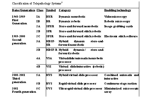
The table shows various generations of telepathology systems.3

DYNAMIC TELEPATHOLOGY (Real Time Telepathology)1,4,5

Is a telecommunication procedure for viewing live images or a sequence of images at real time. It includes remote operation of a microscope and examination of slides from a distance. The system has three requirements:

  1. A robotic microscopic which can be operated remotely.

  2. A workstation which enables the pathologist to remotely operate the microscope.

  3. Adequate and fast telecommunication links.

Advantages

  1. In dynamic telepathology there is no possibility of bias or sampling error. It therefore, presents a perfect scenario with the ability of the remote pathologist  (Teleconsultant) to use the system as a virtual microscope.

  2. The accuracy rate in this system is very high, to the tune of 96%to 100 %.1

Limitations

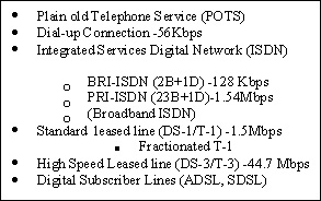
The major limitations are the cost and the necessity of a high bandwidth, to the tune of 1.54 Mb/s for transmission.

STORE AND FORWARD TELEPATHOLOGY (static telepathology)4

Is the practice of pathology at a distance, based on transmission through telecommunication means of still or stationary images from pathology specimens for their corresponding interpretation and diagnosis. It requires necessarily that the referring person is a competent professional pathologist with basic knowledge in pathology informatics. At the receiving centre also there is a pathologist who receives the electronic images and performs the interpretation and diagnosis.

Advantages

  1. Static telepathology is affordable. And therefore, especially useful for small centers with solo pathologists.

  2. A lower level of internet connectivity is required.

Limitations1,4

  1. For a consultant who is used to examining slides in their entirety, it is a suboptimal approach.

  2. The major disadvantage that has been reported is that of sampling error which is due to

- Inability of the Consultant to scan the slide at low power - inability to identify significant

areas at low power.

- Referring pathologists tend to select images which support their own diagnosis.

- Limited number of images may under-represent the complexity of the case.

  1. Errors of interpretation could be related to the consultants' skill and also familiarity with rendering diagnoses on images.

  2. Suboptimal quality of images could also contribute to erroneous diagnoses.

Accuracy of Diagnosis 1,6,7

Accuracy rates of 77% to 99% have been reported by various authors. The studies however, are not comparable, because different methodologies have been employed and the number of participating pathologists in each study is also variable. However, notably, the Arizona International Telemedicine Network has reported a concordance (between the Telepathology diagnosis and glass slide diagnosis) rate of 88.5% and they found a 96.5% concordance for clinically important diagnoses. Weinstein et al have reported a concordance rate of 99%.

HYBRID DYNAMIC STORE AND FORWARD SYSTEMS4

These systems are based on a combination of dynamic and static systems and are more cost-effective.

STATIC TELEPATHOLOGY

Static Telepathology being more relevant in the Indian context, because of lower cost of equipment and telecommunication costs and the lack of broadband linkages4, will be discussed in more detail. The workstation of a static telepathology system is comprised by :
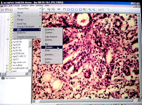
  1. Binocular/Trinocular Microscope

  2. Method for Image Capture (Camera)

  3. Preview monitor

  4. P IV Computer

  5. High resolution monitor

  6. Internet Connectivity

  7. Fax Machine for documentation

Image Acquisition8

Any of the following methods of image capture may be used :

  1. Photographic Images (conventional photography)

  2. Video Camera/Television Images

  3. Digital Camera

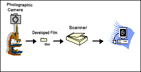
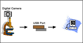
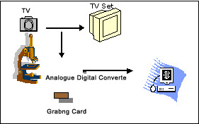
One can use conventional photography using a camera mounted on a microscope, and have the film developed and take prints of images, if that is the only method of image capture available and one does not want to invest further. In this case of course, one would end up spending extra time in having the film developed. One could also use analogue cameras or television cameras to capture images which have been used in the past for display purposes. As of today, however, digital cameras are being used increasingly since they are easy and more convenient to use and are available at increasingly economical prices. Both analogue and digital cameras capture images in the wave form (analogue images) using charged couple devices (CCDs), commonly known as microchips.

The important thing is that images have to be in the digital format in order to permit transfer to a computer, which is necessary to permit transmission of images using internet. While photographic images can be digitized using a scanner, images obtained from an analogue camera are converted into the digital format using analogue to digital converters (ADCs), digital cameras have inbuilt ADCs and therefore, give digital images immediately.

Digital Camera 1,8

Digital cameras available these day are either single-chip cameras or three chip cameras, meaning thereby that they have either a single microchip or three chips inside. A single chip camera offers one third the luminance resolution, because the same CCD accommodates the three primary colours in alteration, whereas a three chip camera has three identical CCDs (each handling a single primary colour) and offers full resolution of an image, which is closest to the true image, this however, is almost triple the cost and there is also a proportionate increase in file size (as explained below).

It is recommended that a single chip camera with a resolution of 3 to 5 megapixels gives optimum images for routine static telepathology purposes.

Digital Imaging 1,8

Digital images are either vector images or raster images. For Telepathology purposes, raster images (bitmaps) are use and will therefore, be discussed.

  1. Vector images

  2. Raster images (Bitmaps ) are an array of pixels or dots. Pixel (picture element or dot) is the smallest unit of digital images and it is assigned a tonal value (which in a bitonal image could be 0 or 1) which is stored in a reproducible sequence by the computer.

  1. 1Bit = 0/1

  2. 1Byte = 8 bits

  3. 1Kilobyte = 1024 Bytes

  4. 1Megbyte = 1000 Kilobytes

  5. 1 Gigabyte = 1000 Megabytes

  6. 1Tertrabyte = 1000 Gigabytes

Colours and Resolution

  1. 1 bit colour image

- 2 (21) colours in a pixel

  1. 8 bit colour image (1 chip camera)

- 256 (28) colours in a pixel (1 byte)

  1. 24 bit colour image (3 chip camera)

- 16.8 (224 ) million colours in a pixel (3 bytes, one each for a primary colour).

Work by Doolittle et al9 has suggested that an 8 bit colour image is optimal for routine static telepathology purposes. The gold standard however, remains 24 bit colour image.

Resolution is in direct relationship with the amount of pixels per unit of longitude

- the larger the no. of pixels captured per image, the higher the resolution.

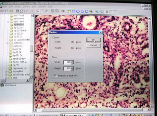
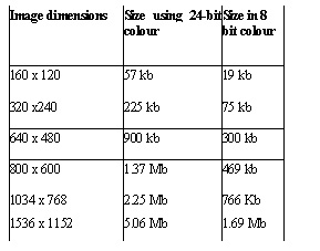
= number of dots per inch (dpi) or pixels per inch (ppi).

File Size = width in pixels x height in

pixels x bit depth in bits

eg : 640 x 480 x 8 = 2457600bits/8

(1byte=8bits) = 307200 bytes

= 300Kb (1kb=1024 bytes)

Recommended Size for transmission

  1. 800 x 600 pixels(475 kb)

  2. 1024 x 750 pixels (768 Kb)


Image Compression

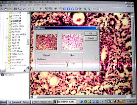
Since large sized images come in the way of easy transmission, it is recommended that images of = 1 Megapixel (1MB) should be compressed. Image compression could be

lossy (20:1 or 30:1 or more, meaning thereby that significant data is lost in compression, or lossless (2:1). One of the more popular formats is the Joint Photographic Experts Group (JPEG), which is generally available with most currently available software. This is a lossy format. Other formats available are Graphics Interchange Format (GIF), Portable Network

Graphics (PNG), Tagged Image File Format (TIFF), Wavelet. However, these formats lack standardization and each one has its own applications. By and large the software that is supplied with the digital camera usually has a mode for resizing the images and compressing them. The figure shows the window for resizing in Olympus CAMEDIA software. Of late, the DICOM standards do apply to pathology imaging too.10

Processing And Optimization Of Images

Images stored in the memory/hard drive of the computer and packed in different formats can be improved or optimized. The contrast, brightness, tone of color, sharpness, size etc. using special programs such as Adobe PhotoShop, PhotoSuite, Picture It, etc. However, for diagnostic Telepathology purposes, the authors do not recommend any changes in the images after they have been captured. It is recommended that since digital cameras generally have software which allow prior changes, all adjustments should be made prior to capturing the images rather than later, for the fear that unintended artifacts may be introduced later. Image optimization should however, be practiced for improving images, when used for presentation, publication etc. The Telepathology Unit of Shree Krishna Hospital uses Olympus C4040 Zoom digital camera with the CAMEDIA MASTER software for opitimization of images.

TELEPATHOLOGY CONSULTANTS

Once imaging has been completed, the next step is to transmit the images to a Telepathology consultant. In static telepathology, it is of utmost important that there is a certain level of confidence and faith between the referring pathologist, the level of communication should also be good. This would help overcome the element of subjectivity to a large extent. Our centre has a collaboration with Tata Memorial Hospital, Mumbai and we therefore, preferentially refer our cases to this centre. There are however, a number of websites which offer Static Telepathology Services, some are free whereas, others charge for their services.

  1. Tata Memorial Hospital

  2. Telepathologyindia.com

  3. UICC

  4. Armed Force Institute of Pathology

  5. Arizona Telemedicine Network

  6. Telepathology.com

Etc.

TELECOMMUNICATION LINKS 11

The advent of internet has brought about the current revolution in information technology and also therefore, medical informatics. For routine static Telepathology, a Plain Old Telephone Service (POTS) generally suffices, which is the usual dial-up connection which transmits data at 56Kb per second. This is the one mode which is easy available to most people and would especially apply to a pathologist in a remote area. Of course, the current availability of better connectivity in cities and digital subscriber lines coming in, data can be transmitted at a much faster rate. The table broadly shows various modes of obtaining internet connectivity with increasing rate of data transmission and therefore, faster speeds at which data could be transmitted. Satellite (V-SAT) is now gaining increasing popularity for effective data transmission and is also being used for the practice of telepathology.

TRANSMISSION OF IMAGES IN STATIC TELEPATHOLOGY1

  1. Images may transmitted as attachments in e-mail attachments, however this permits only a two-communication and therefore, messages would not be accessible to all.

  2. Using File Transfer Protocols (FTP) one can post images on the server which can then be accessed by others

  3. Web Pages permit access to the images by multiple responders and are generally available as websites. This mode permits active interactions amongst groups since the images can be accessed by anyone who wishes to do so.

  4. Telepathology Software Programmes have also been designed to transmit images.

THE INDIAN SCENARIO

Telepathology is relatively new to India. A lot of efforts were put into making it popular with the launch of the website Telepathologyindia.com with prominent pathologists from all over the country on its panel. Up to 93 cases have been analyzed and rendered a telepathology diagnosis by the pathologists of Tata Memorial Hospital, Mumbai. Their rural centre being the Nargis Dutt Memorial Cancer Hospital at Barshi which is 70 kms from Solapur district and 500 kms from Mumbai. Desai et al 12,13,14 concluded:

  1. Concordant / clinically useful diagnosis was rendered in 93.93% of cases.

  2. Major diagnostic discrepancy was observed in 6.06% of cases.

  3. The diagnosis was deferred in only 1 case.

  4. Images were of good and diagnosable quality in 89% of cases.

  5. Turn around time: 8 hrs in 32% of cases 1-3 days in 47% of cases (week-ends).

  6. Static telepathology has the potential to achieve major efficiencies in the diagnostic process and permits specialist support in clinical management.

  7. Store and forward method is economical and is a "one time" investment.

TELEPATHOLOGY UNIT : SHREE KRISHNA HOSPITAL

The Experience So Far

The first Telepathology Unit in the state of Gujarat was inaugurated in November 2002 at the Shree Krishna Hospital, Karamsad. The Unit was started in collaboration with Tata Memorial Hospital, Mumbai.The unit became functional in March 2002. It is equipped with an Olympus CX 41 research microscope, Olympus CAMEDIA digital camera model C 4040 Zoom with CAMEDIA MASTER software a preview monitor and PIV computer with internet connectivity. Karamsad is a village in Anand district. Geographically speaking, the hospital is at an approximate distance of 80 kilometers from Ahemdabad and 45 kilometers from Vadodara. Mumbai is at distance of 420 kilometers. The initial hurdles were related to lack of training and experience not only on part of the pathologist but also personnel supplying the equipment. Not only that, being situated in a rural area, the hospital had a dial-up connection (POTS), at that time. The year 2002, saw 3 cases being submitted for Telepathology consultation, to various centers, using an e-mail account with Indiatimes.com. However, in these three cases, the results were not encouraging and the cases invited only 'deferred diagnosis'. In fact, the cases, though they had been mailed from the account, were not received at all at one of the websites! The turn around time ran into days. In March 2003, the hospital acquired a fractionated leased line, with a bandwidth of 128 kbps. In the mean time, the pathologists at the centre also acquired better imaging skills with increasing familiarity with the software. 19 cases were submitted in the year, with a turnaround time of few hours to 3 days. There were occasions when transmission of images was difficult due to technical reasons. In 2004,till the time of this presentation, 34 cases have been submitted for a Telepathology diagnosis to Tata Memorial Hospital, with a turn around time of a few hours to 1 day in majority of the cases. Cases are sent as e-mail messages with attached images and requisition form. A substantial number of cytology cases have been submitted for diagnosis. The data is being analyzed.

THE INDIAN SCENARIO : PECULIARITIES

Indian conditions present their own peculiarities. They start with poor quality of preparation of basic material, which in fact comes in the way of routine histopathology practice. The basic requirement for the practice of Telepathology is good quality of prepared slides and a microscope with reasonably good optics. In our experience, this does come in the way of obtaining a diagnosis, when cases have to be deferred due to suboptimal quality of sections. Inadequate and inconsistent internet connectivity also comes in the way of an early diagnosis. There have been times when the server is down and it is impossible to transmit cases for technical reasons.

Utility

Is Static Telepathology useful ?

The Shree Krishna Hospital is a rural hospital with a full-fledged cancer centre. There is a young team of pathologists at the department of Pathology. There are occasions when the histopathology diagnosis does not match the clinical diagnosis, and it is desirable to have a second opinion. Many a time, a diagnosis of cancer is itself evoke a request for a second opinion by the patient. When a case is submitted for Telepathology consultation in such circumstances and the report is in agreement with that of the local pathologist, it is highly rewarding - it builds the confidence of the pathologist in himself and also of the clinician in the pathologist and of the patient in the institution. Needless to say, these patients generally stay on with the institution for further management.

There are of course, occasions when a diagnosis is difficult, under these circumstances Static Telepathology is an educational tool for the department.

The practice of Static Telepathology at the Shree Krishna Hospital has helped provide a rapid and expert diagnosis across geographic borders from the premier cancer centre of country at no extra cost rendering an economy of time and resources, which the rural poor of the region could certainly do with.

CONCLUSION

Practiced correctly, Static Telepathology is a useful tool for rendering an early, expert diagnosis (primary or second opinion) rendering substantial economy of time and resources and can prove to be a service, especially to cancer patients in rural areas, by saving them the psychological trauma of having to run from pillar to post for obtaining a diagnosis.

REFERENCES

  1. Leong F Joel W M, GrahamAK, Galun T, McGeeJ O'D. Telepathology: clinical utility and methodology. Recent Advances in Histopathology 18. eds Lowe DG, Underwood JCE. Churchill Livingstone, Edinburgh, 217-239, 1999.

  2. Wells CA, C Sawter. Telepathology: a diagnostic tool for the millennium? J Pathol 2000:191:1-7.

  3. Weinstein RS, Descour MR, Liang C, Bhattacharya AK et al. Telepathology overview: from concept to implementation. Hum. Pathol. 2001, 32, 1283-1299.

  4. Weinstein RS, Bhattacharya AK, Graham AR, Davis JR. Telepathology: A ten-year progress report. Hum Pathol 1997 Jan:28(1):1-7.

  5. Dunn BE, Almagro UA, Choi HY,Sheth NA, Arnold JS, Recla DL, Krupinski EA, Graham AR, Weinstein RS. Dynamic -Robotic telepathology: Department of Veterans Affairs feasibility study. Hum Pathol 1997 Jan:28(1):8-12.

  6. Halliday BE, Bhattacharya AK, Graham AR, Davis JR, Leavitt SA, Nagle RB, McLaughlin WJ, Rivas RA, Martinez R, Krupinski EA, Weinstein RS. Diagnostic Accuracy of an International static-imaging telepathology consultation service. Hum Pathol 1997 Jan:28(1):17-21.

  7. Eusebi V, Foschini L, Erde S, Rosai J. Transcontinental consults in surgical pathology via the Internet.Hum Pathol. 1997 (Jan):28:13-16.

  8. http://www.kodak.com

  9. Doolittle MH, Doolittle KW, Winleman Z, Weinberg DS.Color images in telepathology: how many colors do we need? Hum Pathol. 1997 (Jan):28:36-41.

  10. http://www.medical.nema.org

  11. http://www.ibilce.unesp.br/courseware/datacomm/dc_019.htm.

  12. Desai S, Ghosh TK, Chinoy R, Mohan A, Dinshaw KA. Telepathology at Tata Memorial Hospital, Mumbai and Barshi, a rural centre in Maharashtra. Natl Med J India 2002:15:363-4.

  13. Desai S, Ghosh TK, Chinoy R, Mohan A, Dinshaw KA. Telepathology at the doorstep of a village. Nuclear India 2002: 36:9-10.

  14. Desai S, Patil R, Chinoy R, Kothari A, Ghosh TK, Chavan M, Mohan A, Nene BM, Dinshaw KA. Experience with telepathology at a tertiary cancer centre and a rural cancer hospital. Natl Med J India 2004:17:1,17-19.

Next        Back